
Health and Money
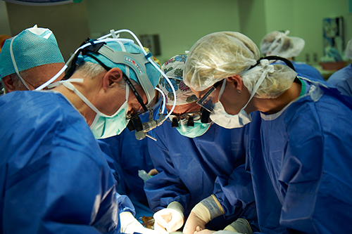
Group of surgeons at work operating in surgical theatre
A routine ultrasound in the 17th week of Charmon’s pregnancy showed the fetus’ abdomen was very distended. Further tests showed there was no fluid in the amniotic sac. Since the amniotic fluid is primarily urine from the fetus, that meant that the baby’s bladder was blocked. If the condition was not corrected, the baby would die.
One week after the ultrasound, Dr. Armando Fuentes at Florida Hospital in Orlando placed a tiny catheter, or shunt, into the fetus’ bladder. This allowed the urine to drain from his body into the amniotic sac, allowing the pregnancy to continue. William was born 11 weeks later, premature and weighing only 2 lbs., 12 oz., but alive.
Fetal surgery, like that performed on William, is one of the newest fields of medicine, one of the most exciting, and one of the most controversial.
“The operations allow for therapy to overcome diagnosis,” explains Dr. Michael Foley of Phoenix Perinatal Associates. He is one of the small group of doctors throughout the country performing fetal surgery. “In the past, we might know that a fetus had an abnormality or serious defect, but we could do nothing until after the baby was born. Now we are finding ways to get into the womb safely and are finding different, innovative treatments to address those conditions.”
Most fetal surgeries are performed on babies with “lethal anomalies.” These are conditions which, if left untreated, will kill the fetus before it is born, like the blocked bladder. Other conditions doctors treat with surgery are separating acardiac twins (only one of the twins has a heart and is trying to provide blood for both fetuses), diaphragmatic hernias (a tear in the thin sheet of tissue separating the heart and lungs from the abdomen), and excessive buildup of fluid in the chest cavity (which pushes the heart of one side and causes heart failure).
The early 1970s saw a near-revolution in new medical equipment and techniques. As microscopic instruments, CAT scans, heart-lung supports, ultrasound, and new drugs were developed for other areas of medicine, perinatal specialists – those who deal with pre-birth conditions – began looking at ways to use them on their patients.
Early operations in the mid-1970s involved ‘open fetal surgery’ – actually opening the womb, removing the fetus while still keeping it attached to the umbilical cord, then replacing the fetus, and reclosing the mother’s womb.
“It was very radical,” explains Dr. Fuentes. “If you find the defects and can repair them early enough, the babies don’t die. But the fetus is non-viable under 20 weeks.” That was a major problem, since doctors preferred doing the surgery earlier than that, if they could.
Even if the surgery was successful, there were other complications. “The mother might go into premature labor, and 15 years ago there were not many good medications then to stop labor. We would have to deliver the baby early, and then would have trouble closing the womb.”
Doctors performed the surgeries when they felt there was a chance of saving the baby, since if they did nothing, it would certainly die. But as less-invasive procedures evolved, the open fetal surgeries largely ended. New ways to combine ultrasound and laparoscopes (a thin tube equipped with an eyepiece, lenses, and light source. Some procedures can be performed using instruments threaded through the tube.), more micro-sized instruments, and advances in laser surgery give doctors many options for new treatments.
But the wonders of the technology must be balanced by the moral aspect of what some people consider tampering with a life that’s barely started. In and out of the medical community, there is a major debate over the ethics of fetal surgery. Many of the babies who are born after fetal surgery spend months in the hospital and face years, perhaps a lifetime, of medical care. Infants with kidney problems like William must sometimes be on dialysis from birth and almost always need a kidney transplant when they are as young as two years old. Mental retardation and other physical and developmental problems are common. It is emotionally exhausting for the families and the medical providers. Treating the children represents an enormous financial outlay for the families, insurance companies, hospitals, and society.
. “There are so many variables to determine,” says Dr. Carl Weiner, Professor and Chair of Obstetrics, Gynecology and Reproductive Services at the University of Maryland Medical Center. “We can save their lives, but is it a kindness?” says Dr. Weiner. “We need to step back and study the implications of the outcomes of the surgeries.”
That’s a sentiment echoed by his colleagues. All fetal surgery centers have ethics panels which review each potential surgery on a case-by-case basis. It’s a difficult, sometimes heartbreaking procedure. Everyone involved – physicians, nurses, technicians, social workers, and clerks- want to see parents go home carrying their newborn child, but they know that is not always possible.
It’s their responsibility to decide which cases are good candidates for surgery. While each hospital has its own specifics, the ethics panels share general guidelines. The first is that the condition which brought the baby to them is the only physical problem. There are tests to make sure the baby’s chromosomes are normal and that there are no other birth defects. If those conditions exist, the panel will not approve the surgery. Unfortunately, some unborn babies are just too broken to be fixed. In those cases, the panel usually recommends terminating the pregnancy before the mother’s health is jeopardized.
If the case passes the first review, then the panel takes a hard look at the family. Even if the team is optimistic about the outcome of the surgery, it’s a difficult, high-stress situation that will last beyond the pregnancy, quite possibly for the life of the child and its family.
“We look for a lot of family support,” Dr. Fuentes says of his panel. “Not just the parents, but their extended families, as well. The parents see a genetic counselor and meet with a psycho-social counselor. They have to understand that this is not 100% guaranteed.”
At Phoenix, Dr. Foley involves the parents in the analysis of their case. “It helps if we take the steps together. We set the guidelines up front, so everyone knows what the risks and the decision points are. Communicating with the parents is very important. In every case, Mom is the baby’s advocate. She wants to do whatever she can to help and protect her baby. Dad is Mom’s advocate. He doesn’t want to do anything that will put her at risk.”
Until recently, the surgeries were confined to conditions that were fatal. But in the last two years, doctors have begun operating on fetuses with serious, but non-fatal, conditions.
The Vanderbilt University Medial Center made headlines this summer when it announced doctors performed brain surgery on a fetus that was diagnoses with hydrocephalus, or water on the brain. A shunt was placed in the brain to allow fluid to drain into the amniotic sac. Without the operation, the baby probably would have been born with a severely enlarged and deformed head filled with fluid and almost no brain function. Most children with hydrocephalus die before their tenth birthday. The infant showed no signs of the defect after birth, but it will be six months to a year before doctors will know if there was brain damage.
In the last two years, about 60 operations have been performed on fetuses diagnosed with Spinal Bifida. The most common debilitating birth defect, it is found in one out of every 1,000 pregnancies in the U.S. The spine does not completely develop, leaving an opening in the back and the spinal cord exposed. Babies born with Spinal Bifida suffer disabilities like paralysis and weakness in their lower limbs, urinary and bowel dysfunction, skeletal deformation, mental retardation, and hydrocephalus.
The surgery performed on these fetuses differs from the others in two ways. First, Spinal Bifida is not a fatal condition. The fetus would probably live and be born after a full-term pregnancy. Secondly, these operations took doctors back to performing open-womb surgery – removing the baby from the mother in order to perform the operation, then returning it, and hoping the mother continues to a full-term pregnancy.
The debate over the procedure started with the first operation. The question is whether the doctors are giving in to the temptation to see a problem and fix it without understanding how one problem treated in isolation might affect the baby’s overall health.
“If we have a chance to lessen the extent of injury, why wouldn’t we do that?” asks Dr. Joseph Bruner, Director of Fetal Diagnosis and Therapy at Vanderbilt University, a pioneer in the technique.
“We don’t truly understand the effect of a birth defect on the baby,” counters Dr. Weiner. “There’s a risk in repairing the fetuses if you don’t know what the risks are post-natally.” A recognized condition may effect the fetus in ways doctors do not realize. Fixing one problem may not improve, or may ever worsen, others. In cases of non-fatal birth defects, the best thing for the baby might be to let it be born with an untreated defect, for which effective post-natal treatment exists.
The answer lies in whether the operation really does improve the child’s condition. “It’s somewhat encouraging,” says Dr. Foley. “As the children are getting older, we can assess the development of their motor skills and brain. There seems to be some reasons for enthusiasm. But a truly just decision of whether this should be done requires matching a control group of children who did not have the operation and comparing their conditions and development.” It may turn out that babies treated in utero for non-fatal conditions are not noticeably healthier than those born with the same defects without treatment.
“We must ask ourselves why we are branching out to non-lethal conditions,” agrees Dr. Fuentes.
But branching out is inevitable. “Different, innovative treatments are going to be the future,” says Dr. Foley. He’s proof of his own prediction. He’s developed a tiny spring wire grasping device that is inserted into the womb through a needle and catches the umbilical cord. Using ultrasound, he can move the fetus to a better position when he performs surgery.
The debates don’t mean much to parents like Charmon Shuford. All she cares about is that she has her son, and any ordeal she and her husband endure for him is worth it. During the 13 weeks her son was in the hospital after he was born, she spent every day at his side. “I’d come home to eat dinner. My husband would get home from work and then we’d go back at night and stay until about eleven o’clock. We were only home to sleep.”
Now he is home, letting the Shufords have a chance at acting like normal parents. They know William will probably need a kidney transplant as soon as he old enough and strong enough to have one. If his kidney function drops below a critical level before then, he will have to begin dialysis.
“We’re playing a waiting game,” she says, while William laughs in the background. “And we pray a lot. But every time something comes up and we get through it, we decide we’ve cleared one more hurdle.”